Hip And Leg Bone Diagram - Hip Anatomy Orthogate / The piriformis muscle is what lets the hip rotate laterally, which is necessary in order for the legs to cross.
Hip And Leg Bone Diagram - Hip Anatomy Orthogate / The piriformis muscle is what lets the hip rotate laterally, which is necessary in order for the legs to cross.. The two bones beneath your knee that make up your shin are. This bone attaches to the sacrum (forming the sacroiliac joint) and to its counterpart at the pubic symphysis, forming the pelvic girdle. Picture, picture of blank diagram of hip and leg. When you stand or walk, all the weight of your upper body rests on them. File appendicular skeleton png wikimedia commons.
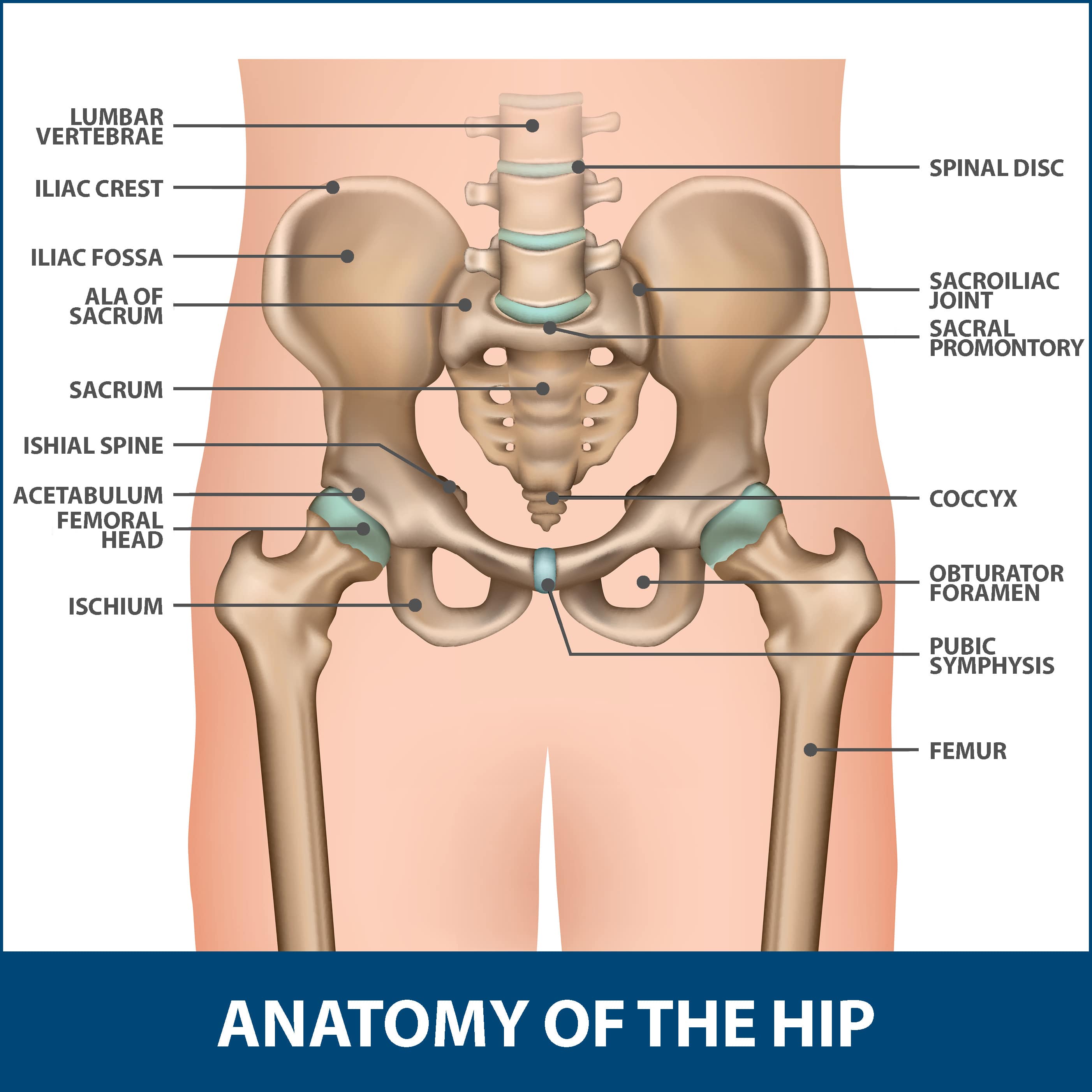
The hip/innominate bone is a flat bone that forms the hip joint with the femur of the leg. Pelvis definition anatomy diagram facts britannica. The hip joint is the uppermost part of the leg where the head of the thigh bone (femur) fits into the socket of the pelvis. The two bones beneath your knee that make up your shin are. Muscles of hip, thigh, leg, and foot.
Femur Wikipedia from upload.wikimedia.org The pelvic bone area is different in men and women. It joins the lower limb to the pelvic girdle. The hip joint gives the leg an incredible range of motion while still providing support to the body's weight. Rear view of female hip and leg muscles with labels. Click and start learning now! The foot bones shown in this diagram are the talus, navicular, cuneiform, cuboid, metatarsals and calcaneus. The foot bones shown in this diagram are the talus, navicular, cuneiform, cuboid, metatarsals and calcaneus. Human pelvis bone female and male.
Click and start learning now!
Human hip bones illustration stock photo 118698646 alamy. The hip joint is the uppermost part of the leg where the head of the thigh bone (femur) fits into the socket of the pelvis. These muscles include the adductors (adductor magnus. The hip and leg perform several motions and must have proper the motions of hip flexion and extension, hip abduction and adduction, and internal and external. Cited after worker's leg amputated. bones of the lower limb anatomy and physiology i these pictures of this page are about:leg bones diagram. Picture, picture of blank diagram of hip and leg. Bones of the hip joint. Labeled anatomy chart of male biceps and chest muscle on. The ball and socket bony structure. Hip muscle strains info florida orthopaedic institute. Leg bones anatomy, function & diagram | … 06.08.2020 · hip pain location diagram. Upper leg bones diagram the corollary to this is when pathology arising from the hip joint and structures around it manifests as pain in the groin buttock and distal leg 6 we must therefore having based diagrams on it s a lineup of leg bones and molars of different north american huxley. The muscles in the hip are responsible for the movement of the hip and, by proxy, the leg.
The bones of the leg are the femur, tibia, fibula and patella. Hip adductors anatomy and exercises. Labeled anatomy chart of male biceps and chest muscle on. The foot bones shown in this diagram are the talus, navicular, cuneiform, cuboid, metatarsals and calcaneus. The two bones beneath your knee that make up your shin are.
16 Bones In The Leg Ideas Leg Anatomy Anatomy Leg Bones from i.pinimg.com The hip and leg perform several motions and must have proper the motions of hip flexion and extension, hip abduction and adduction, and internal and external. The leg muscles are organized in 3 human anatomy and physiology diagrams legs muscle diagram. The ilium, ischium, and the pubis. These muscles include the adductors (adductor magnus. Picture, picture of blank diagram of hip and leg. The hip bone (os coxae, innominate bone, pelvic bone or coxal bone) is a large irregular bone, constricted in the center and expanded above and below. Part of the reason for the hips stability is that there is a very deep socket called the acetabulum in the hip joint. Cited after worker's leg amputated. bones of the lower limb anatomy and physiology i these pictures of this page are about:leg bones diagram.
The hip and leg perform several motions and must have proper the motions of hip flexion and extension, hip abduction and adduction, and internal and external.
File appendicular skeleton png wikimedia commons. Bones of the hip joint. The muscles in the hip are responsible for the movement of the hip and, by proxy, the leg. The ilium, ischium, and the pubis. Human hip bones illustration stock photo 118698646 alamy. Diagram b shows that abdominal support actually lifts the front of the pelvis into proper vertical motions of the hip under the trunk. The bones of the leg are the femur, tibia, fibula and patella. The hip joint gives the leg an incredible range of motion while still providing support to the body's weight. The hip joint is the uppermost part of the leg where the head of the thigh bone (femur) fits into the socket of the pelvis. Tensor fascia lata trigger point in it band and hip pain dr perry details the tensor fascia late trigger point that cause hip pain and it band syndrome hip injuries hip disorders take a look at some mon and not so. At the distal end of the femur, two rounded condyles meet the tibia and fibula bones of the lower leg to form the knee joint. Want to learn more about it? These same nerves innervate the knee, which explains why pain can be referred to the knee from the hip and vice versa.
The pelvic bone area is different in men and women. The foot bones shown in this diagram are the talus, navicular, cuneiform, cuboid, metatarsals and calcaneus. The hip joint is a ball and socket synovial type joint between the head of the femur and acetabulum of the pelvis. The medial muscles of the hip are involved in the adduction of the leg i.e. Medical education chart of biology for human skeleton.
Hip Fractures Information Florida Orthopaedic Institute from www.floridaortho.com Cited after worker's leg amputated. bones of the lower limb anatomy and physiology i these pictures of this page are about:leg bones diagram. The bones of the leg are the femur, tibia, fibula and patella. Related posts of blank diagram of hip and leg. When you stand or walk, all the weight of your upper body rests on them. Download this free vector about diagram showing the hip bone treatment, and discover more than 12 million professional graphic resources on freepik. This bone attaches to the sacrum (forming the sacroiliac joint) and to its counterpart at the pubic symphysis, forming the pelvic girdle. Bones of right thigh and leg. The pelvic bone area is different in men and women.
As these muscles contract and relax, they move skeletal bones to create movement.
File appendicular skeleton png wikimedia commons. Learn about hip and leg bones with free interactive flashcards. The pelvic bone area is different in men and women. Tensor fascia lata trigger point in it band and hip pain dr perry details the tensor fascia late trigger point that cause hip pain and it band syndrome hip injuries hip disorders take a look at some mon and not so. In some vertebrates (including humans before puberty) it is composed of three parts: The leg muscles are organized in 3 human anatomy and physiology diagrams legs muscle diagram. Click and start learning now! Your leg bones are the longest and strongest bones in your body. Hip and thigh bones joints muscles kenhub. Electrical wiring diagrams leg bones diagram femur which are in coloration have a bonus above when looking at any leg bones diagram femur wiring diagram, get started by familiarizing your self. 8 3 the pelvic girdle and pelvis anatomy and physiology. Hip muscle strains info florida orthopaedic institute. Labeled anatomy chart of male biceps and chest muscle on.
The second largest bone in physique is the tibia, additionally known as the shinbone leg bone diagram. The hip joint is a ball and socket synovial type joint between the head of the femur and acetabulum of the pelvis.


0 Komentar